


The Parts of a Horse: Essential Knowledge for Every Rider

Could you point to a horse’s hock, stifle, and fetlock without mixing them up? These names can feel like a new language at first, especially when care guides use familiar-sounding words for different joints. Learning the parts of a horse gives you a practical map: you can locate each region and understand its basic role.
It’s common to confuse body areas that look similar or use terms that don’t match human anatomy. You don’t need to memorize every detail to follow horse-care conversations. This guide works from the head and neck through the body and limbs, explaining where key visible regions are and how to tell commonly confused terms apart. With a head-to-tail map and a closer look at each leg, you can describe what you see more clearly and follow anatomy references with greater confidence.
Table of Contents
What Are the Main Parts of a Horse? A Head-to-Tail Overview
Horse anatomy is the study of a horse’s body structure. To get your bearings, follow the outline from head to tail: the head and neck lead into the trunk, followed by the hindquarters, legs, and hooves. This simple map helps you place unfamiliar terms without having to learn every structure at once.
The main external regions are the head, neck, trunk, hindquarters, legs, and hooves. Each includes smaller areas with their own names. This overview focuses on what you can see from the outside; a broader reference such as Equine Anatomy also covers internal structures and body systems.
Where to find the head, neck, trunk, and hindquarters
Start at the head. The poll is at the top of the head, just behind the ears. The muzzle is the front of the face around the nostrils and mouth. The neck joins the head to the body and slopes toward the shoulders.
The shoulder is the region around the top of the forelegs. The withers sit at the top of the shoulders, where the neck meets the back. They provide a useful landmark when describing the horse’s outline. The barrel is the rounded trunk beneath the back, behind the shoulders and in front of the hindquarters. The chest is at the front of the trunk, while the flank is the area between the barrel and hind leg.
At the rear, the croup is the upper surface of the hindquarters near the base of the tail. The tail extends from the back of the body. The legs begin beneath the trunk and hindquarters and end in hooves. Moving from one region to the next makes it easier to follow anatomy descriptions and locate each term on the horse.
What Do a Horse’s Legs and Hooves Include?
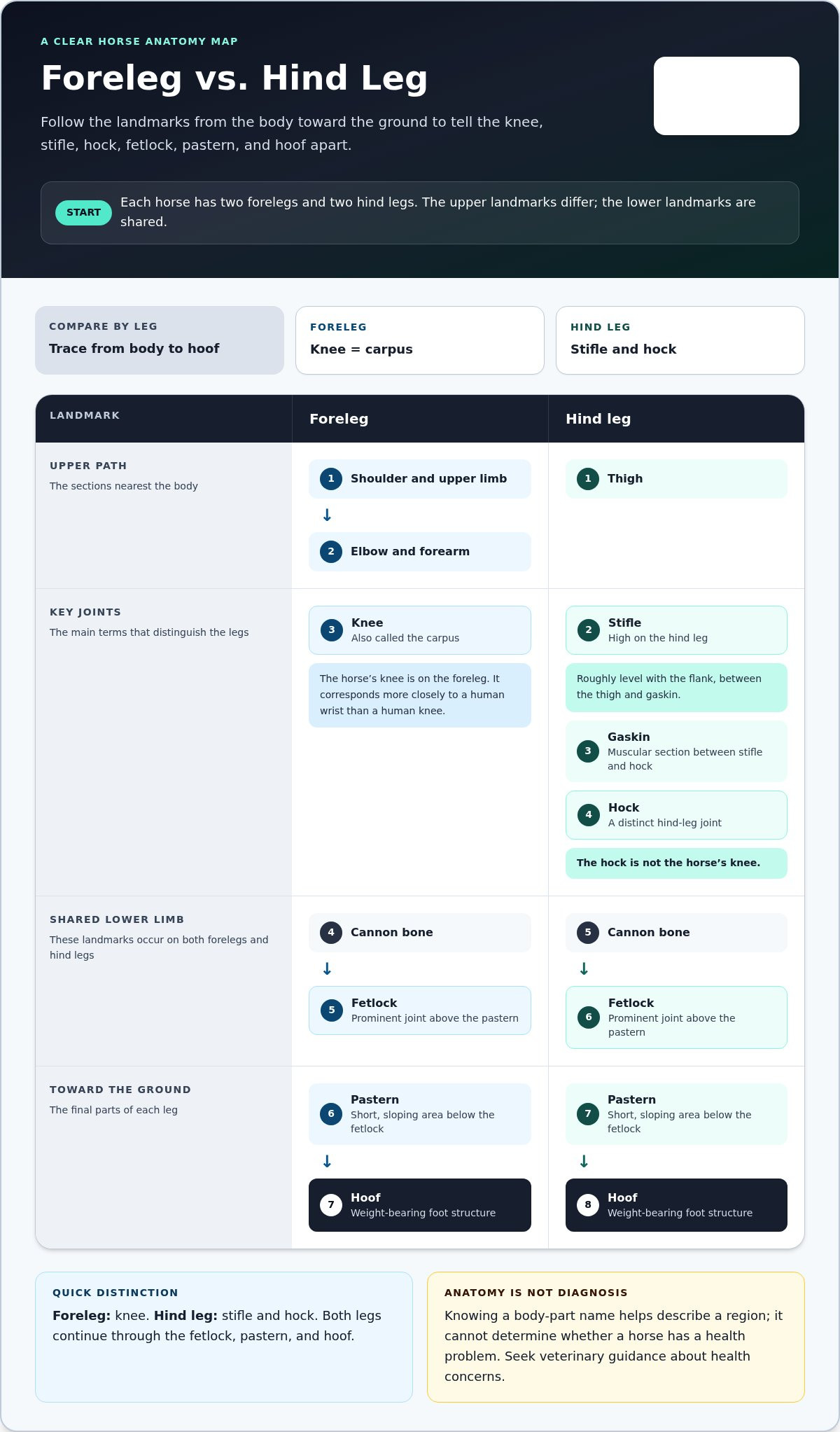
A horse has two forelegs and two hind legs. Some landmarks are shared, especially on the lower limbs, but the upper sections differ. Follow each leg from the body toward the ground to place the terms accurately. A useful first distinction is that the horse’s knee is on the foreleg, while the stifle and hock are on the hind leg.
How to distinguish the knee, hock, fetlock, pastern, and hoof
Use these simple sequences as a text-based leg map:
Foreleg: shoulder and upper limb → elbow and forearm → knee, also called the carpus → cannon bone → fetlock → pastern → hoof.
Hind leg: thigh → stifle → gaskin → hock → cannon bone → fetlock → pastern → hoof.
The horse’s “knee” is the carpus, a joint in the foreleg that corresponds more closely to a human wrist than a human knee. The stifle is a large joint high on the hind leg, roughly level with the flank, between the thigh and gaskin. The gaskin is the muscular section between the stifle and hock. Farther down the hind leg, the hock is a distinct joint; it is not the same as the horse’s knee.
On both forelegs and hind legs, the fetlock is the prominent joint above the pastern. The pastern is the short, sloping area between the fetlock and hoof. Below it, the hoof is the horse’s weight-bearing foot structure. It forms the end of each limb and supports the horse as it stands and moves. Following the leg in order helps you avoid common mix-ups, such as calling a hock a knee or confusing a fetlock with a hoof.
Knowing these names helps you follow care discussions and identify which region someone is describing, but anatomy vocabulary alone can’t determine whether a horse has a health problem. For reliable horse-health information, consult resources such as the American Association of Equine Practitioners (AAEP), and seek veterinary guidance about concerns. Learning the parts of a horse’s legs is a useful foundation for understanding how each limb is described.
How Can You Use Horse Anatomy Terms in Everyday Learning?
Accurate anatomy terms make equine learning easier to follow. If a lesson or care resource refers to the withers, fetlock, or croup, knowing where each area is helps you picture the instruction and describe what you notice. Naming a region is a useful observation, but it isn’t a diagnosis.
Build a simple, repeatable horse body-parts vocabulary
Study a labeled diagram from head to tail, then review the legs separately. A short, repeatable routine helps the names stick:
Point to each visible region and say its name aloud, starting at the poll and moving toward the tail.
Trace each leg from the body down, noting where forelegs and hind legs differ and where they share terms.
Check a term’s location on a labeled diagram if you’re unsure, particularly for the stifle, hock, and knee.
Use the terms in a neutral description, such as “the area below the fetlock,” rather than guessing what a change might mean.
Learning the parts of a horse gives you a clearer foundation for reading educational materials and asking focused questions. It supports informed horsemanship, but it doesn’t replace professional assessment. If you’re concerned about a horse’s health, describe what you’ve observed and seek guidance from a veterinarian. Clear, careful language can help keep anatomy learning useful without turning a body-part name into a health conclusion.
Build Confidence Through Clear Anatomy
A head-to-tail map helps you recognize the main parts of a horse. Reviewing the legs separately makes terms such as knee, stifle, hock, fetlock, pastern, and hoof easier to distinguish. Using these names accurately can make equine education materials clearer and help you describe what you observe.
Anatomy vocabulary is a foundation for learning, not a way to diagnose a health concern. If something about a horse’s health worries you, share your observations with a veterinarian. You can build your understanding gradually by learning a few terms, locating them on a diagram, and then noticing how they relate to the surrounding regions.
For a structured next step in foundational horsemanship, explore Texas Equine Education & Horsemanship™’s horsemanship lessons and education-first approach to learning from the ground up.
Frequently Asked Questions
What are the main parts of a horse’s body?
The main parts of a horse are the head, neck, trunk, hindquarters, legs, and hooves. From front to back, the head includes the poll behind the ears and the muzzle around the nose and mouth. The neck joins the head to the body. The trunk is the central body, and the hindquarters form the rear around the croup and tail. Paired legs extend below the body and end in hooves.
What is the difference between a horse’s withers and croup?
The withers are at the top of the shoulders, where the neck meets the back. The croup is farther back, forming the upper surface of the hindquarters near the tail. Both are landmarks along the horse’s back, but they’re at opposite ends of the body: the withers are near the forelegs, while the croup is above the hind legs.
Is a horse’s hock the same as its knee?
No. The hock is a joint on each hind leg, located between the gaskin above and the cannon area below. The horse’s “knee” refers to the carpus, a different joint on the foreleg. The terms name distinct locations, so identifying whether you’re looking at a front or hind limb helps avoid confusion in anatomy discussions.
What are the parts of a horse’s leg called?
From top to bottom, a foreleg includes the shoulder, elbow and forearm, knee (carpus), cannon area, fetlock, pastern, and hoof. A hind leg includes the thigh, stifle, gaskin, hock, cannon area, fetlock, pastern, and hoof. The shared lower-leg terms refer to similar locations, but the upper regions differ. A labeled diagram can help you follow each sequence.


Comments